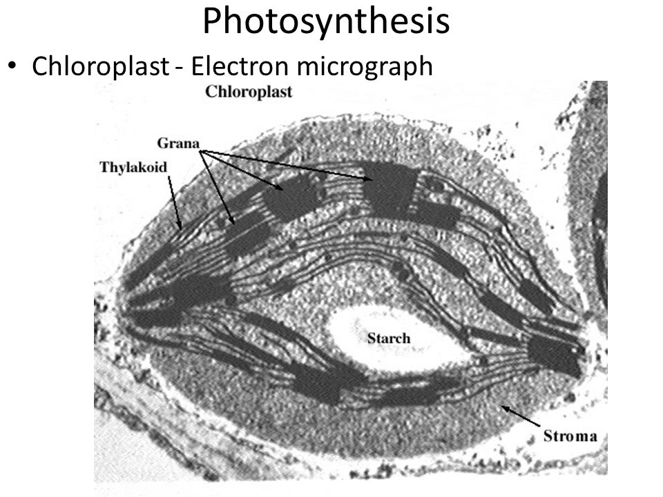
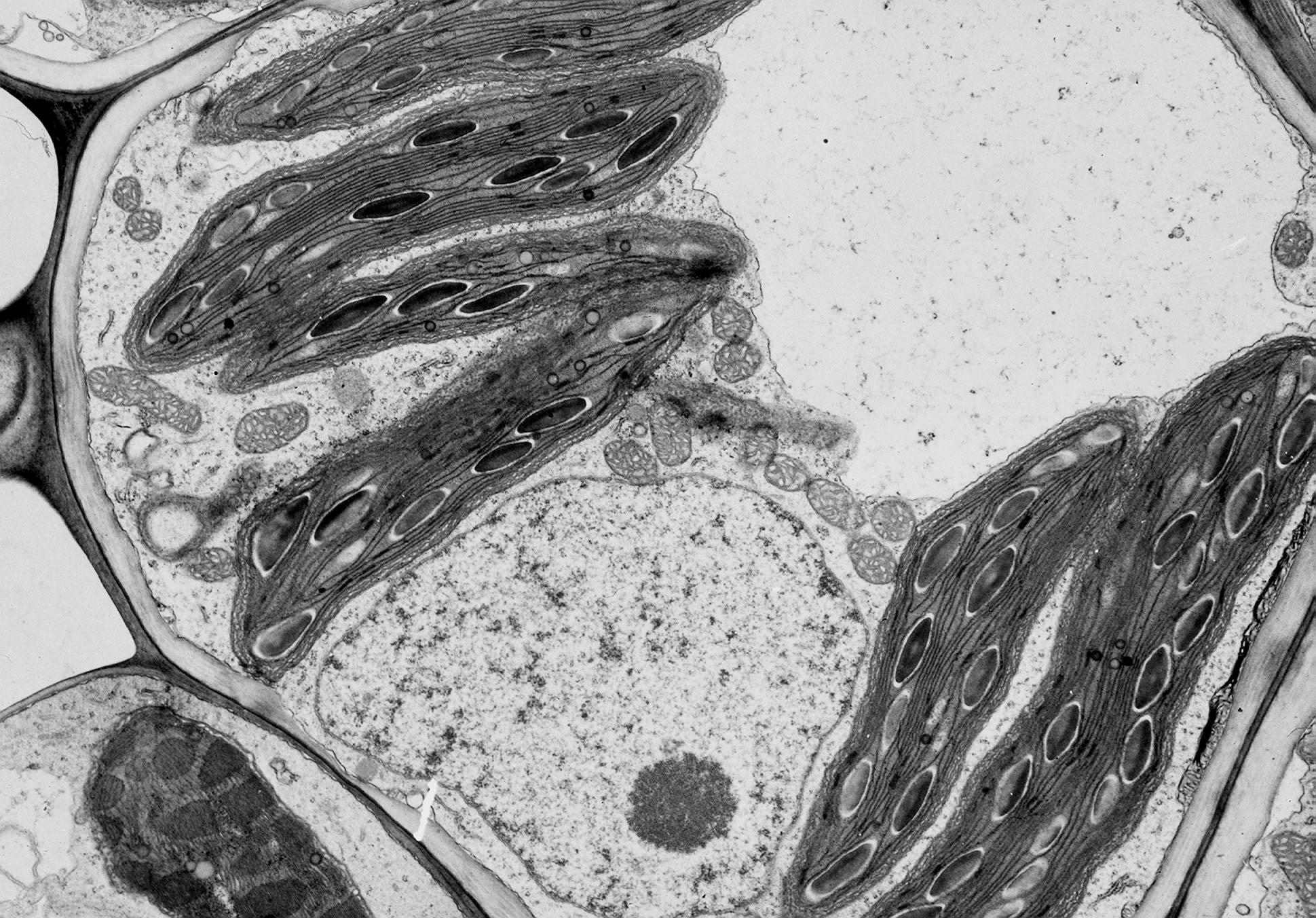
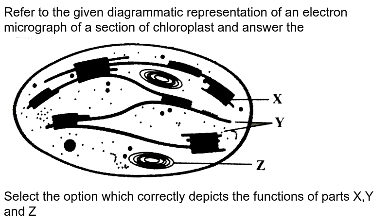
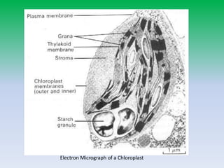
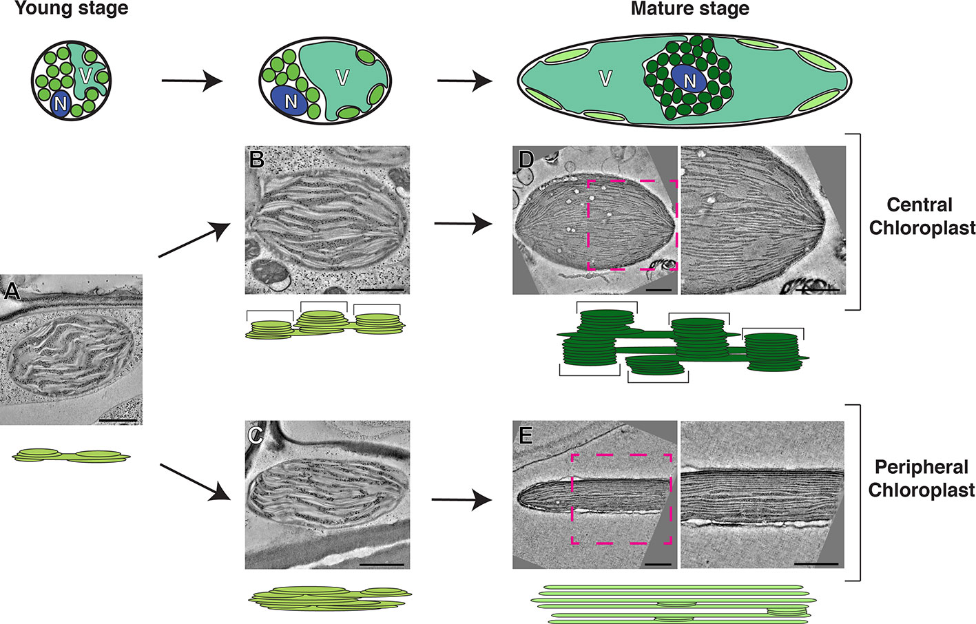
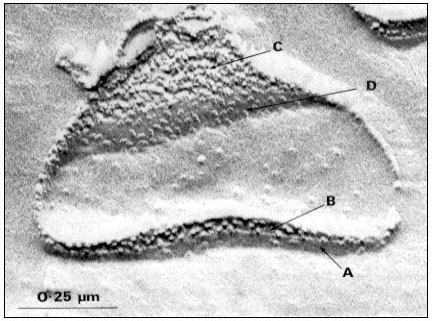
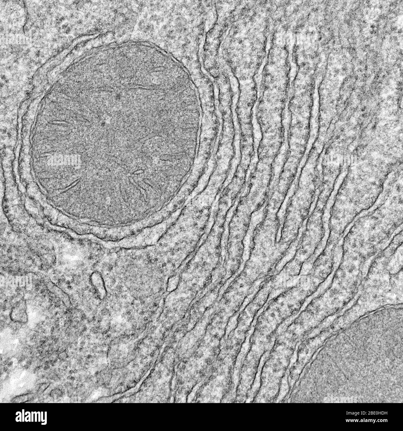
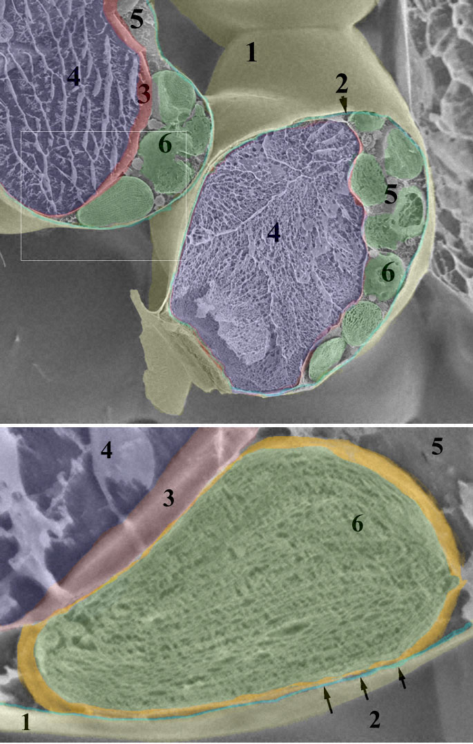
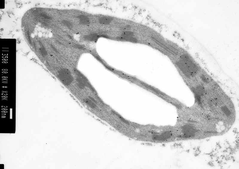
40 labelled electron micrograph of chloroplast
Join LiveJournal Password requirements: 6 to 30 characters long; ASCII characters only (characters found on a standard US keyboard); must contain at least 4 different symbols; Glossary of botanical terms - Wikipedia pl. apices The tip; the point furthest from the point of attachment. aphananthous (of flowers) Inconspicuous or unshowy, as opposed to phaneranthous or showy. aphlebia pl. aphlebiae Imperfect or irregular leaf endings commonly found on ferns and fossils of ferns from the Carboniferous Period. aphyllous Leafless; having no leaves. apical At or on the apex of a structure, usually a shoot, a stem ...
Cambridge International AS & A Level Biology Coursebook … Suppose we want to know the actual length of the labelled chloroplast in this electron micrograph. R ev ie w C op y WORKED EXAMPLE 4 ev ie Pr es s -C To calculate the real or actual size of an object, we can use the same magnification equation. separate points. If the two points cannot be resolved, they will be seen as one point. In practice, resolution is the amount of detail …

Labelled electron micrograph of chloroplast
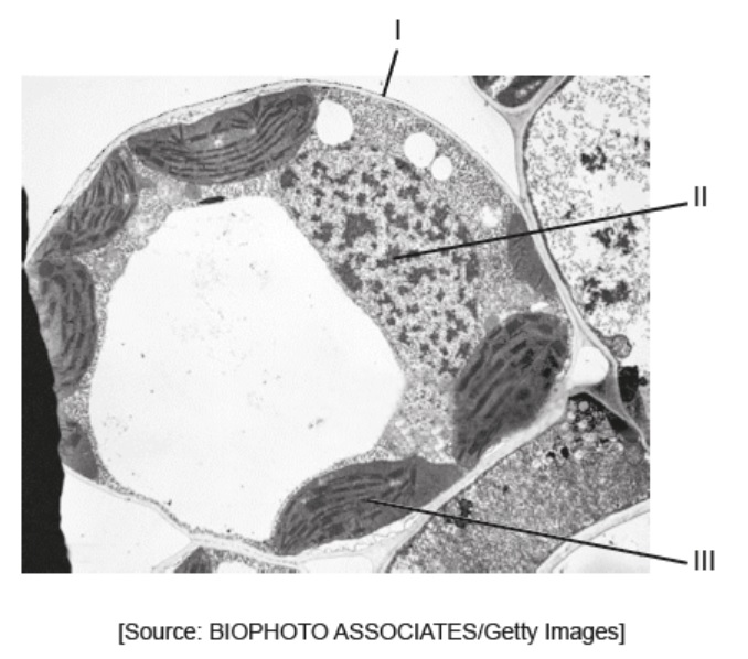
Cambridge IGCSE Biology Coursebook (third edition) - Issuu Jun 09, 2014 · The black spots in the electron micrograph in Figure 2.8 are granules of a carbohydrate called glycogen. This is similar to starch. (Starch is never found in animal cells – they store glycogen ... A Level Biology For OCR A (PDFDrive) | PDF - Scribd “4 Figure 3 Tansmission electron ‘micrograph ofa section through two leaf cells at their junction, Their cell walls run from top centre to lower lft. Anucleus is Seer it kane ng ht Skat gromales 1 Study the two drawings above and state, with. {pale ovais) can be seen in the chloroplast (dark grey, upper left and right). x18 700 ‘mognification reasons, which of them represents the best ... Microscopes - Save My Exams An electron micrograph of the same leaf mesophyll cell at the : same magnification: would show more detail than is shown in Fig. 1.1. (a) Explain why, at the : same magnification, an electron micrograph is able to provide more detail than a light micrograph..... [2] (b) Describe: three additional features that could be seen on an electron micrograph of the leaf mesophyll cell that …
Labelled electron micrograph of chloroplast. Cambridge International AS and A Level Biology ... - Academia.edu BIO1: Maintaining a Balance 1. Most organisms are active in a limited temperature range IDENTIFY THE ROLE OF ENZYMES IN METABOLISM, DESCRIBE THEIR CHEMICAL COMPOSITION AND USE A SIMPLE MODEL TO DESCRIBE THEIR SPECIFICITY ON SUBSTRATES Cambridge IGCSE Biology Third Edition Hodder Education Enter the email address you signed up with and we'll email you a reset link. Glenn Toole - Aqa Biology A Level Student Book-Oxford ... - Scribd More than one electron may be lost or received, for • The loss of an electron leads to the formation of a example the loss of two electrons from a calcium atom positive ion, for example, the loss of an electron forms the calcium ion, Ca 2•.1ons may be made up of more from a hydrogen atom produces a positively charged than one type of atom, for example a sulfate ion … Nelson Biology 12.pdf [30j71j2z320w] - doku.pub Electrons are not included in this number because the mass of an electron is negligible compared with the mass of a proton or neutron. The mass of an atom is therefore determined by the number of protons and neutrons it contains. Table 2 lists the atomic numbers and mass numbers of the most common elements in living organisms. The atomic symbol of an element is sometimes …
Microscopes - Save My Exams An electron micrograph of the same leaf mesophyll cell at the : same magnification: would show more detail than is shown in Fig. 1.1. (a) Explain why, at the : same magnification, an electron micrograph is able to provide more detail than a light micrograph..... [2] (b) Describe: three additional features that could be seen on an electron micrograph of the leaf mesophyll cell that … A Level Biology For OCR A (PDFDrive) | PDF - Scribd “4 Figure 3 Tansmission electron ‘micrograph ofa section through two leaf cells at their junction, Their cell walls run from top centre to lower lft. Anucleus is Seer it kane ng ht Skat gromales 1 Study the two drawings above and state, with. {pale ovais) can be seen in the chloroplast (dark grey, upper left and right). x18 700 ‘mognification reasons, which of them represents the best ... Cambridge IGCSE Biology Coursebook (third edition) - Issuu Jun 09, 2014 · The black spots in the electron micrograph in Figure 2.8 are granules of a carbohydrate called glycogen. This is similar to starch. (Starch is never found in animal cells – they store glycogen ...














Komentar
Posting Komentar